Preparation:
The sample was embedded in resin, cut along two sections, and polished. Surface cleaning with ethanol and N₂ (5 bars) was performed.
A carbon coating by evaporation with a high-purity braid (thickness ~30 nm) was applied to ensure secondary electron conduction in scanning electron microscopy.
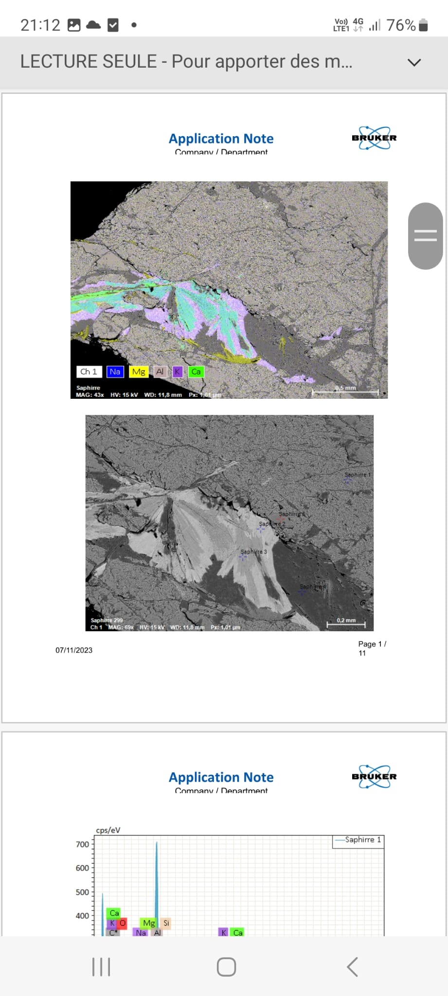
BSE Imaging:
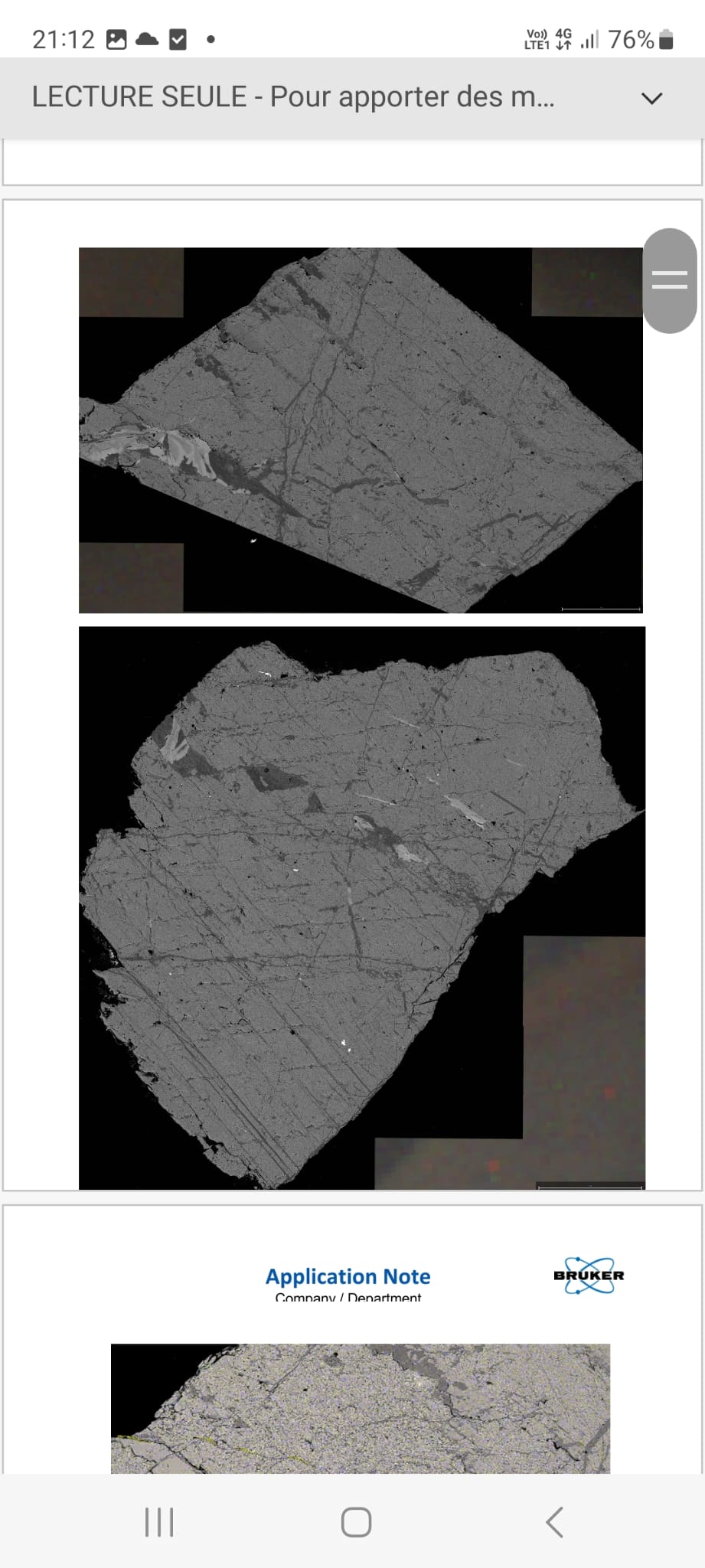
High-resolution mosaics (25 Mpixels) were obtained in backscattered electron imaging (atomic weight contrast) covering both sections, in secondary vacuum (~2 × 10⁻⁵ Pa) (column conditions: 15 keV, spot 4, aperture 30 µm, working distance 10 mm).
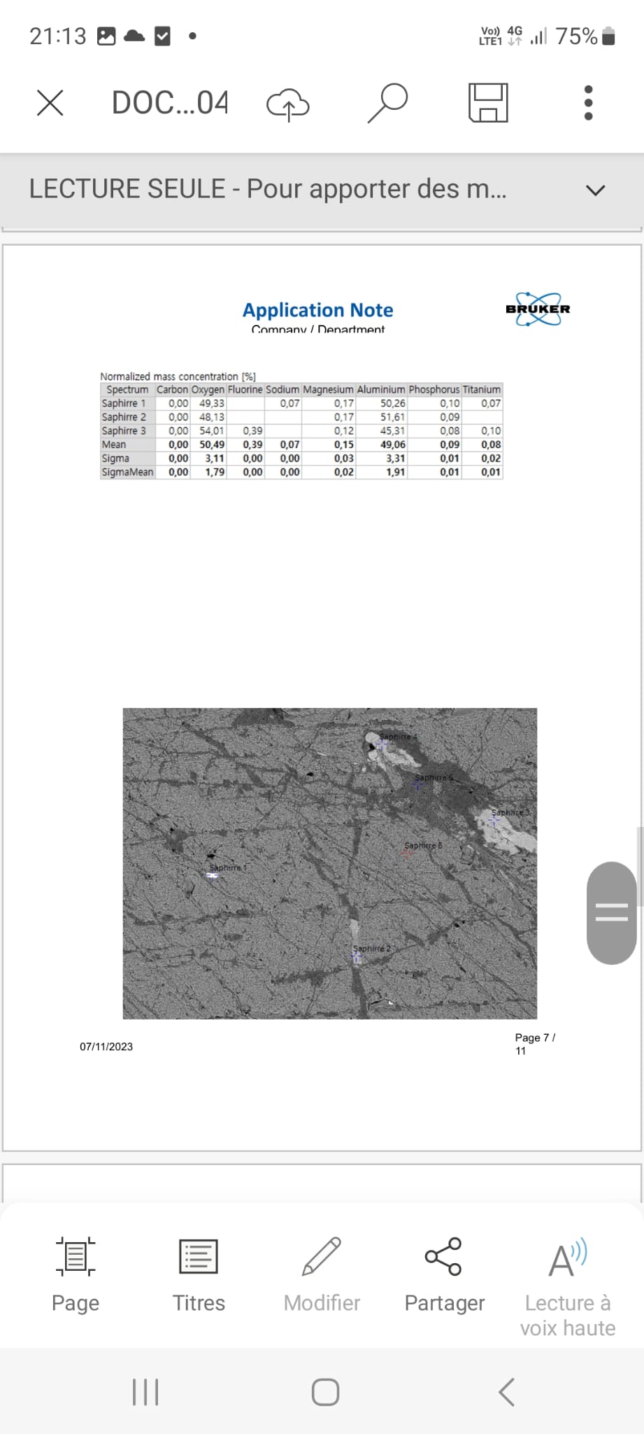
The textural analysis confirms a heterogeneous polyphase sample, showing networks of filled fractures and small automorphic minerals included, in both sections (see mosaics 1 and 2).
EDS Elemental Mapping:
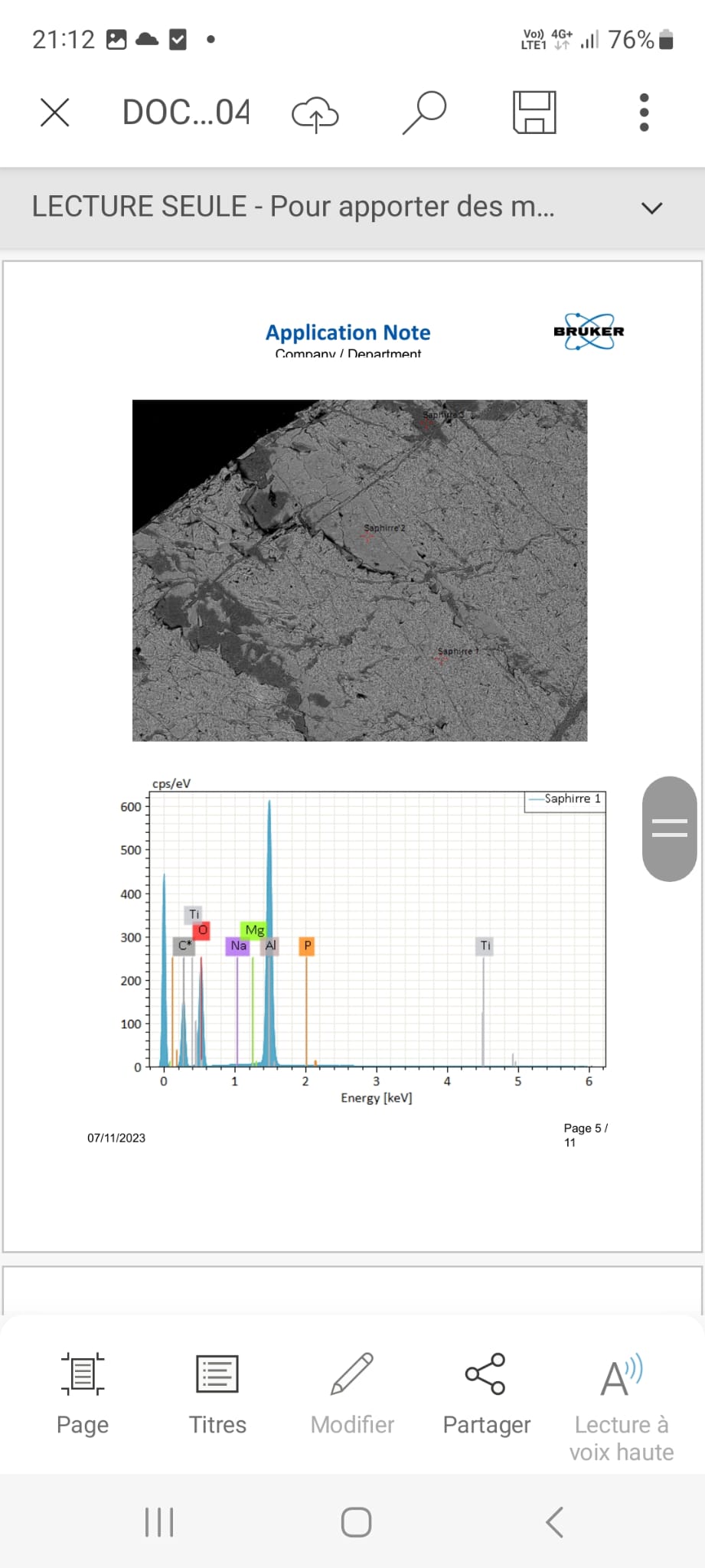
A qualitative elemental mapping was carried out on one section (see mapping), in secondary vacuum (column conditions: 15 keV, spot 5.5, aperture 50 µm, working distance 11.8 mm, EDS time constant: 600 kcps/s), with a resolution of 1500 pixels.
This confirms the aluminum oxide stoichiometry of the main phase, as well as three distinct fillings in a large fracture: two first phases whose texture and composition appear to indicate a phyllosilicate (Phase A rich in Ca and Na, Phase B rich in K), and a filling phase of xenomorphic aluminum oxide, depleted in Al compared to the main phase.
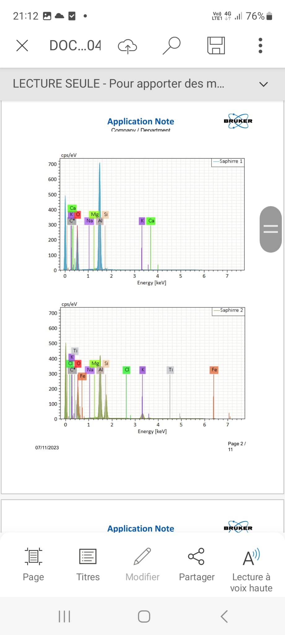
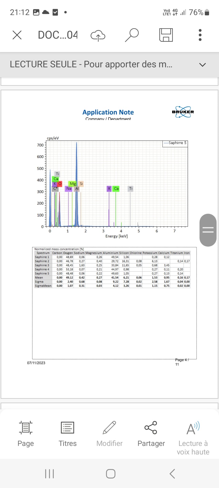
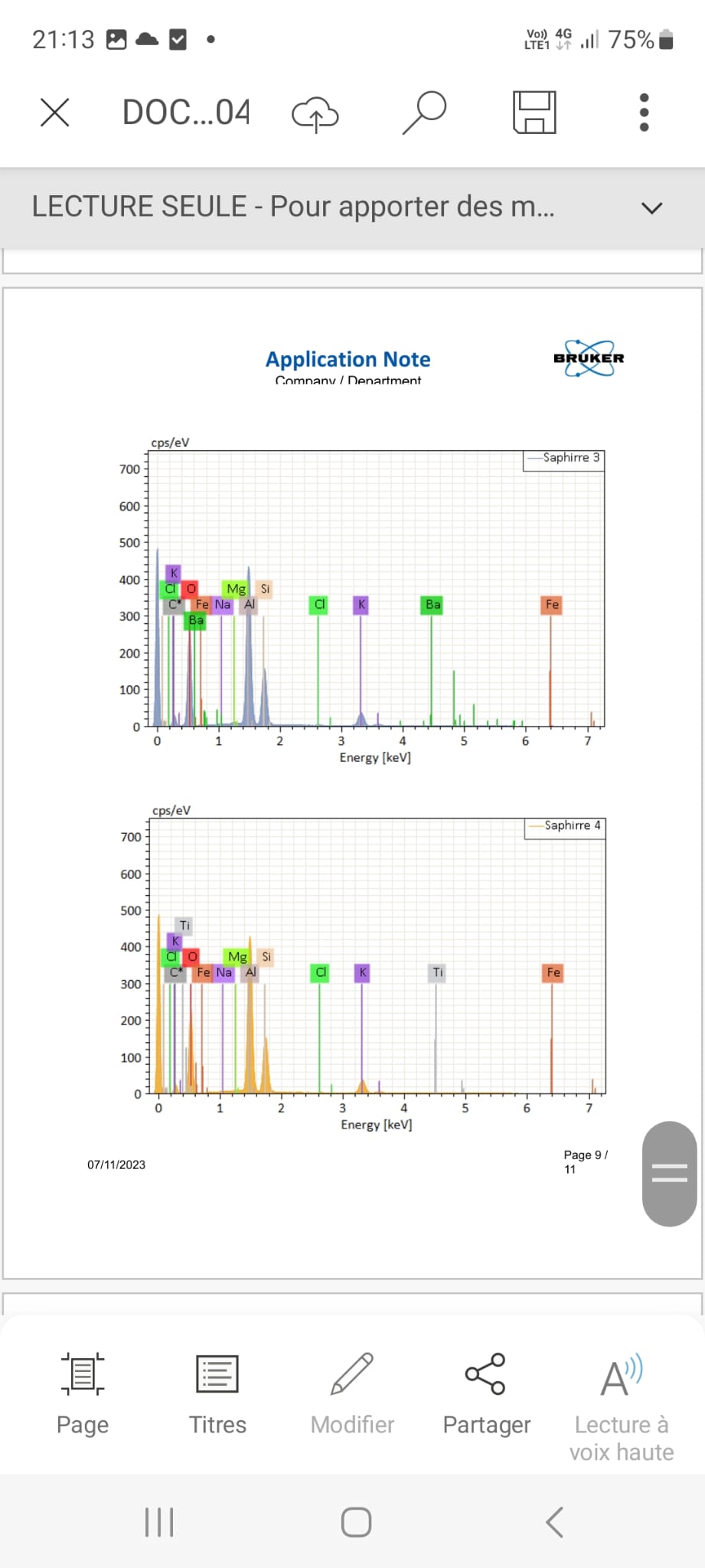
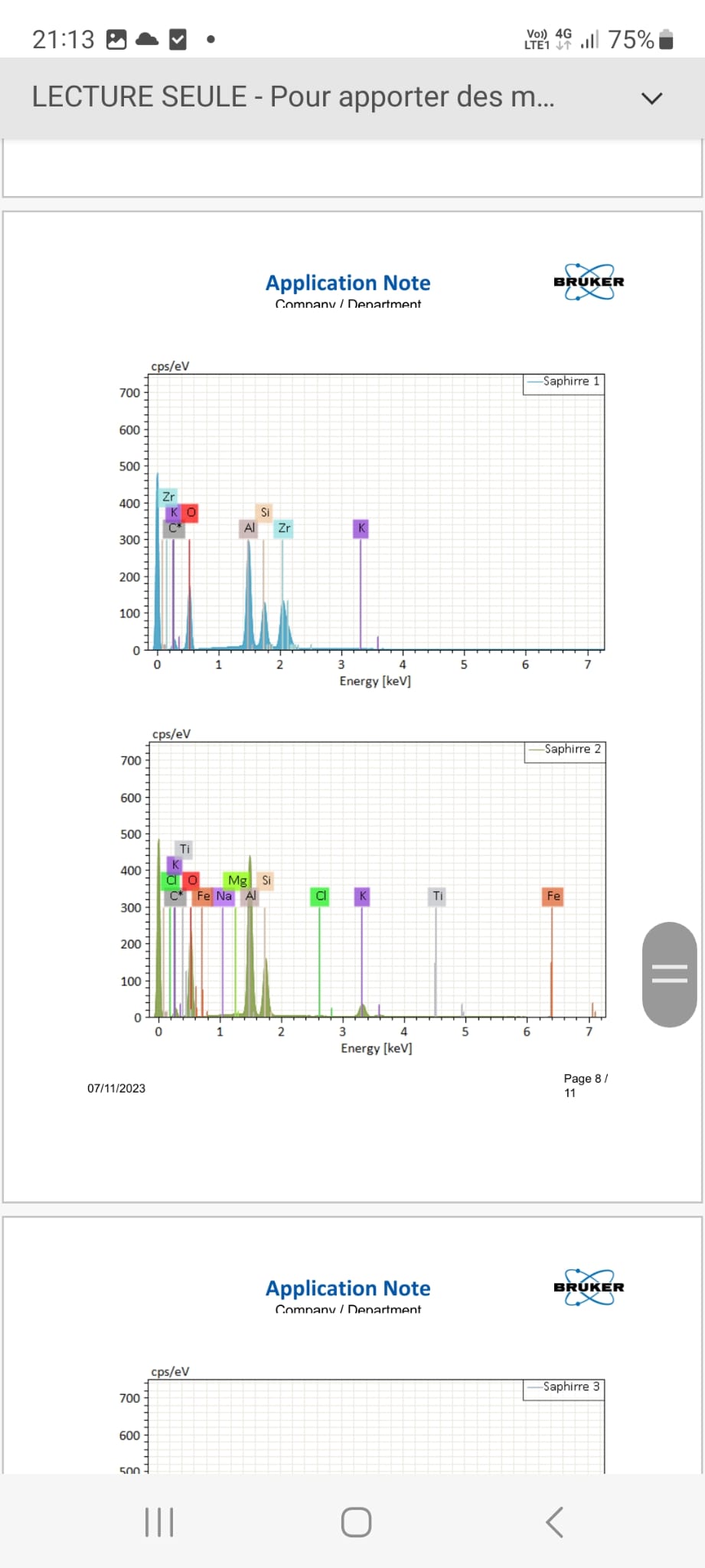
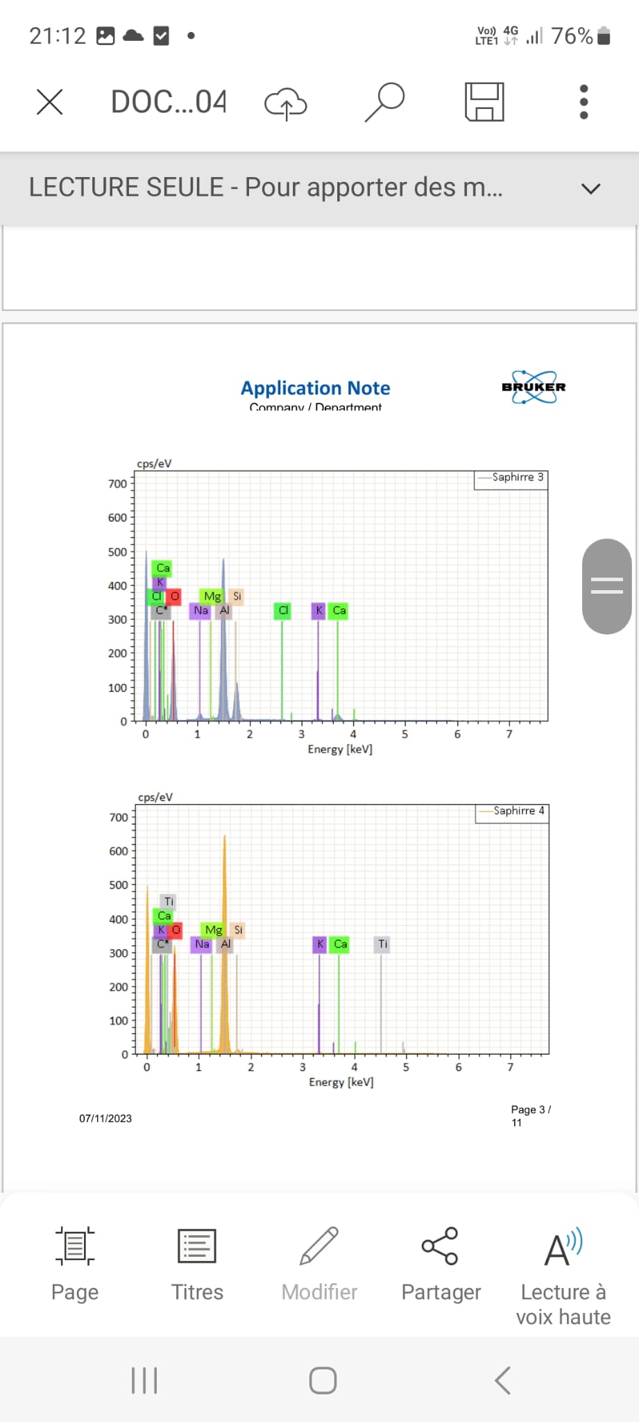
EDS Spot Analyses:
Quantitative spot analyses were carried out in two areas of the first section and one area of the second section, in secondary vacuum (column conditions: 15 keV, spot 5, aperture 30 µm, working distance 11.8 mm, EDS time constant: 130 kcps/s).
The interaction volume is estimated at 1.6 × 2.9 µm (L × W) under these conditions. The atomic weight elements below Na are not detectable in EDS; O is quantified in EDS, especially in this case. Quantifications are performed after Rho/Phi/Z corrections with certified and calibrated standards (MAC CAS) under these analytical conditions.
-
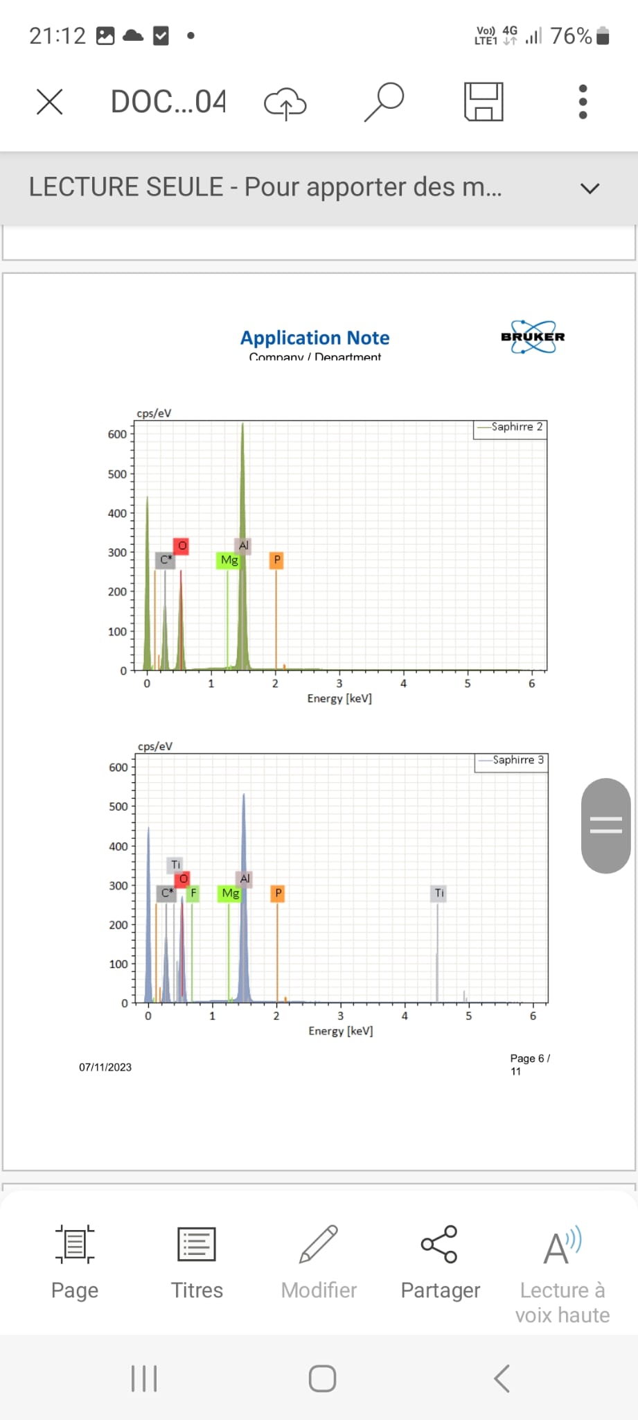
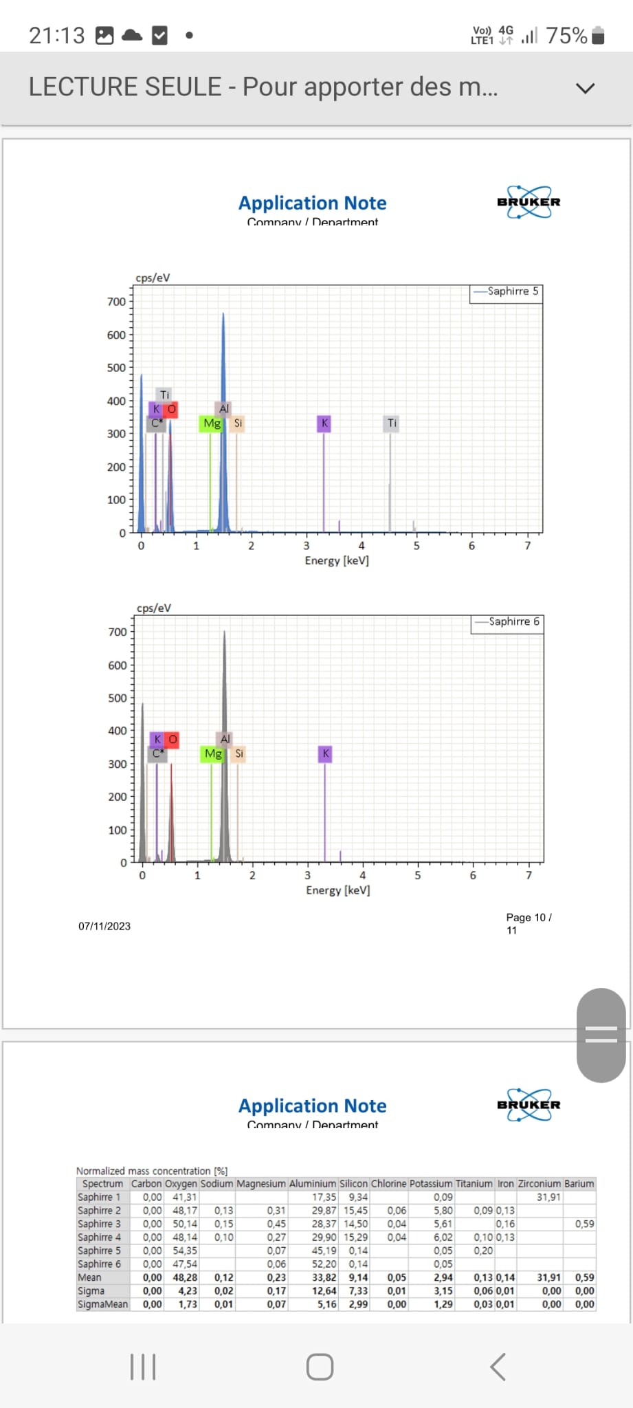
Main phase: Analyses confirm the presence of corundum with stoichiometry ~Al₂O₃, with traces of Ti, Ca, K, and Mg in some areas (Image 1 spectra 1 & 5; Image 2 spectra 1 & 2; Image 3 spectrum 6), consistent with sapphire.
-
Inclusions: Very rare automorphic zircons were detected as inclusions in the main phase (Image 3 spectrum 1).
-
Fracture filling: Filling of numerous fractures/joints in 3 distinct phases seems likely:
-
A = Growth of a calcic-sodic phyllosilicate (micas) automorphic with traces of Mg (Image 1 spectra 3).
-
B = Overgrowth of a second potassic phyllosilicate phase (traces of Fe, Ba, sometimes V) (Image 1 spectra 2 and Image 3 spectra 2, 3, 4) completely filling some fractures and partially others (see mosaic 2).
-
C = Total filling of porosity by a xenomorphic aluminum oxide which does not respect the stoichiometry of corundum (~AlO₂) (Image 1 spectrum 4; Image 2 spectrum 3; Image 3 spectrum 5).