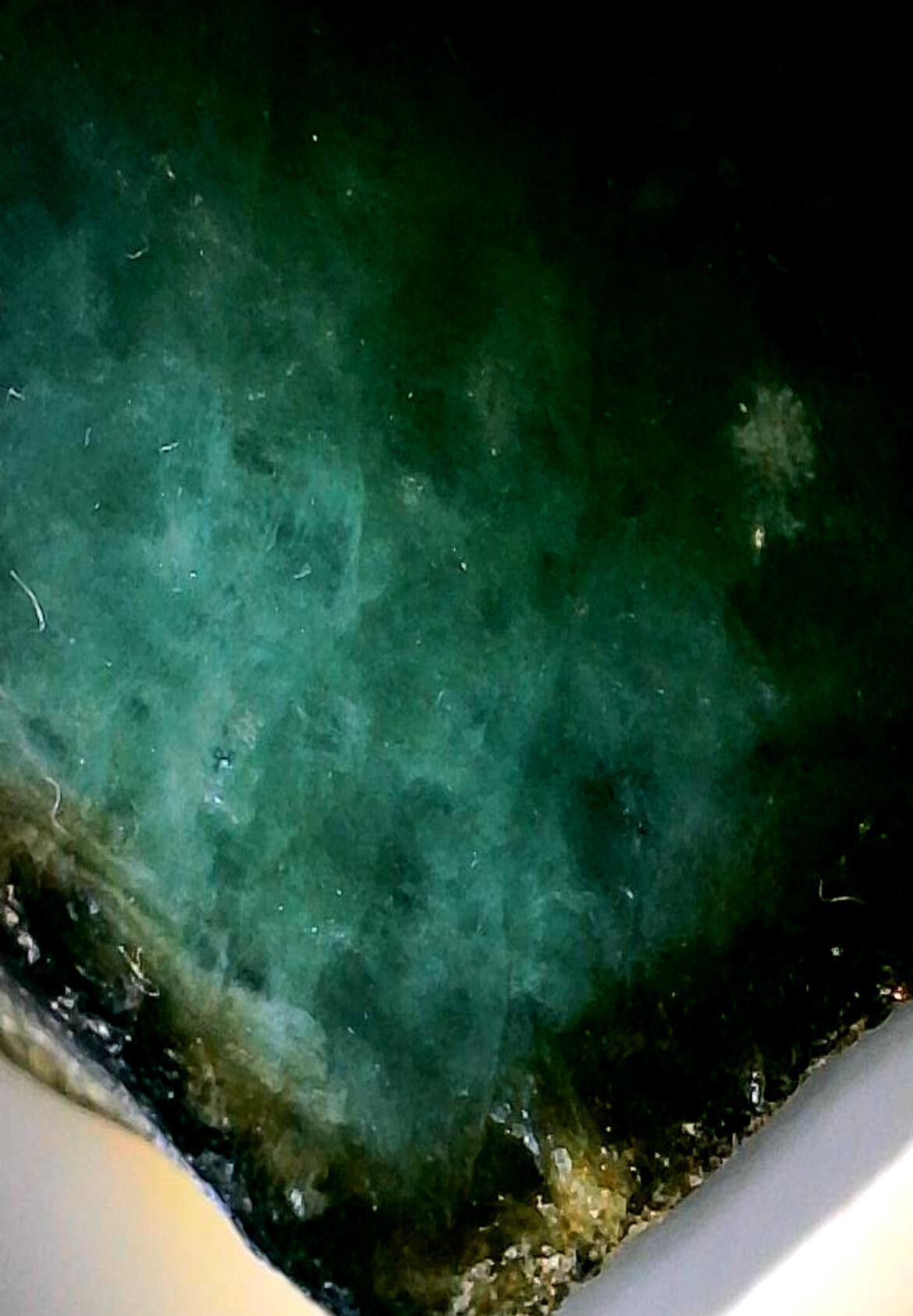
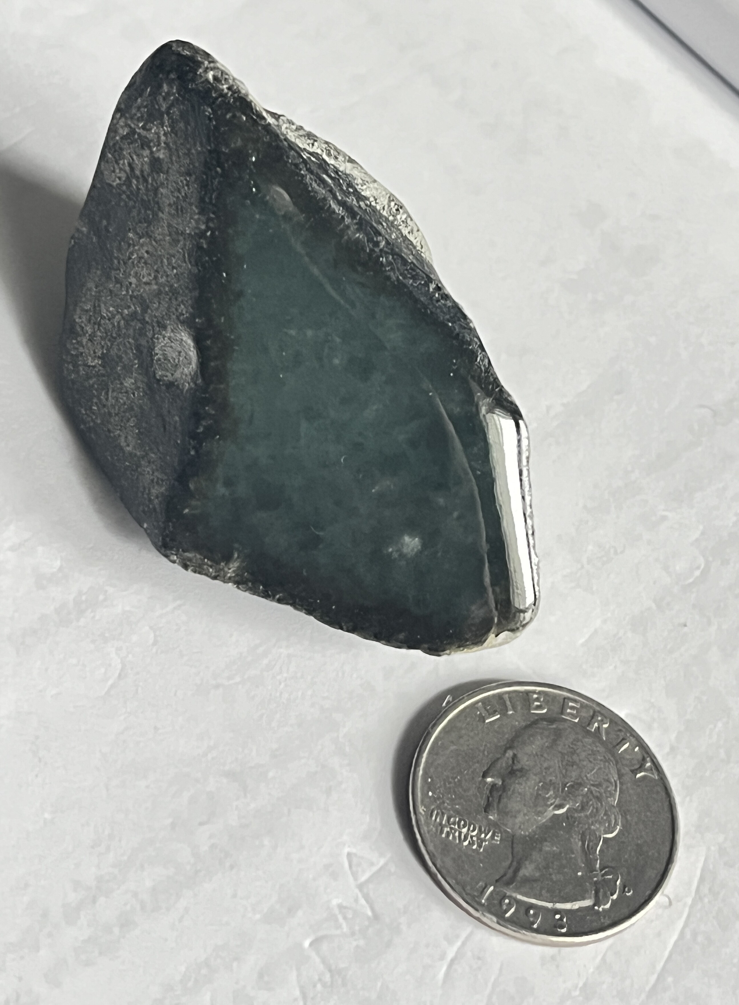
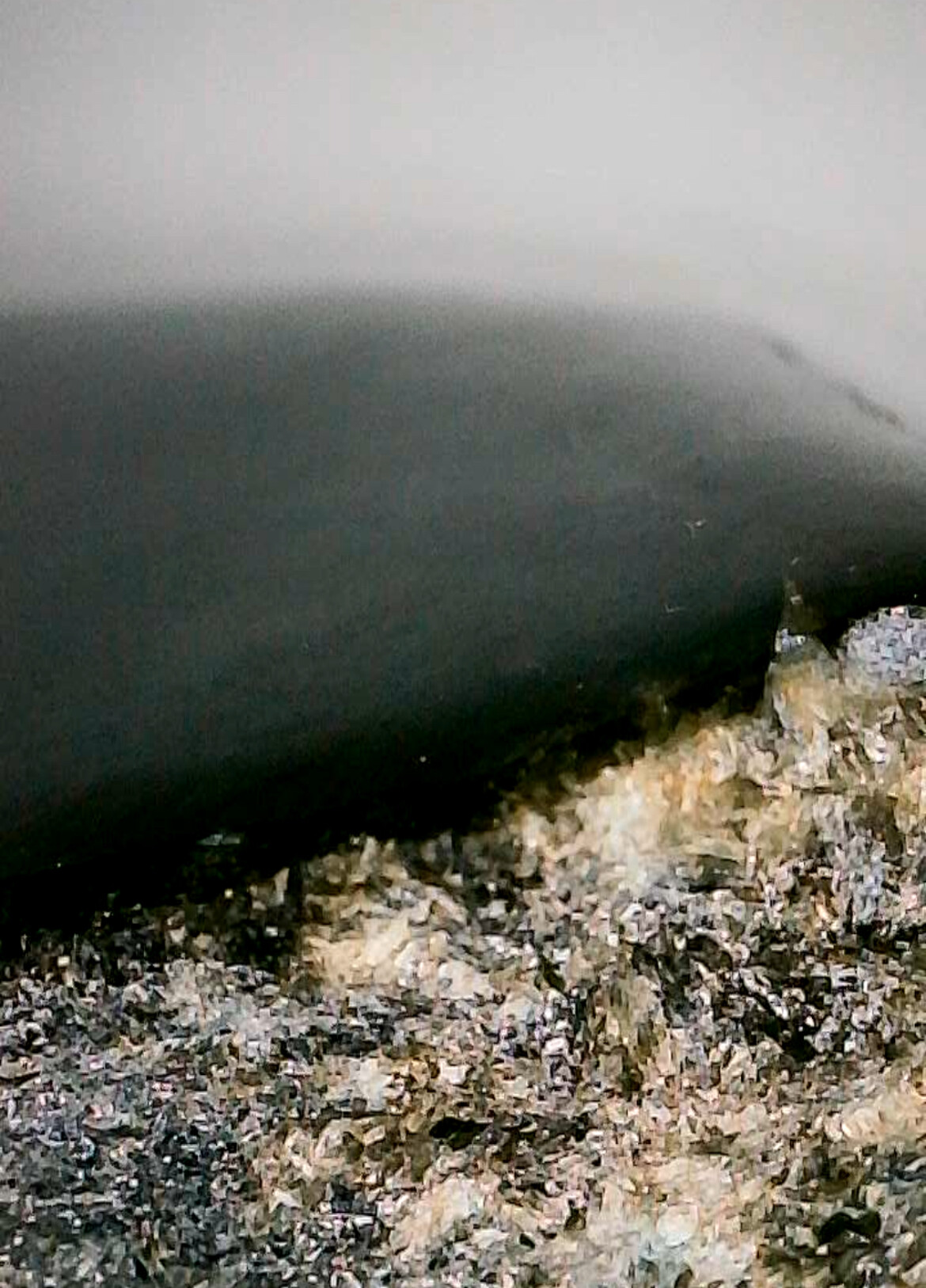
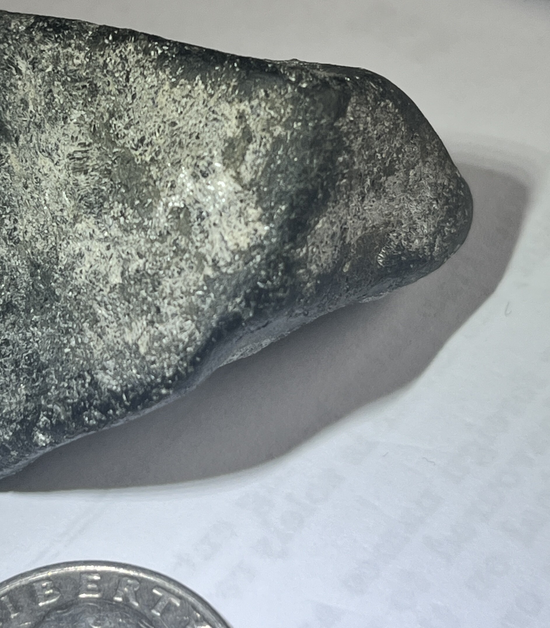
I recently purchased a very interesting (to me) stone. It was found in the Middle East, but who knows where it’s from originally. One side is polished and the other are rough, it was caked in dirt when I got it but upon washing it off, I realized the polished surface is an emerald like green, with inclusion patterns that look to me like they may be emerald. I measured the RI of the "emerald surface and its about 1.59-1.6. It’s also within the emerald Mohs range, per my measurements about 7.5. The host rock def has a mica like sparkle to it, and may be biotite, if my hunch/math is correct.
Here’s the math part: the specific gravity of the whole stone (host plus possible emerald) is around 3.2, which at first had me conclude it wasn’t emerald, but given emerald (if it is) ~2.75, so long as the host mineral is greater than the total SG of the stone, the emerald part may still be emerald.
By my math, assuming biotite mica host (SG 3.3), roughly 82% of the could be that mineral and the remainder (about 18%) could measure 2.75, ie. emerald. Eyeballing it, that seems possible given the surface area of the polished part vs the rest.
It’s a big stone, 372cts total, which would mean 66cts of emerald (hypothetically). I don’t want to break it up but I wonder what others think. I like it either way, so no disappointment if it’s not emerald, but if not, any guesses on what it could be?
Here are some pics.
Would this specimen have come from the Emerald Mining Districts along the Red Sea?
Not sure if a Chelsea Filter will help in this situation, but if you can attempt to observe it through one using light to illuminate the material internally, it could help see if chromium is present.
Dichroism? A dichroscope would help.
Any reaction to UV?
Hi, I truly have no clue where it may have been mined, but it was found most recently in the Middle East. I didn’t know there was a source in Egypt, but looked it up and learned a lot, thanks for the data-point.
This is probably a stupid question, but is a spectroscope and a dithiscope the same thing? I have a cheap version of the former that I have never been able to use effectively (at all, actually), but not the later. Could I test using the UV filter that was included in my low-end refractometer? I’ve never used it before, but if it could work, I’d love to learn how best to measure.
I just checked UV again and I don’t think it fluoresces. The shiny part is reflective, so I see that effect, but I don’t think it is fluorescence.
Thank you!
Dichroscope and spectroscope are different. (It’s a good question…)
The dichroscope can detect pleochroism (at least two different color hues) and helps determine if a stone is double refractive. It uses a set of polarized filters or an optical calcite window to allow the user to observe the effect.
The spectroscope can help identify absorption lines in the visible light spectrum (some can reach into the near IR and/or into the UV bands). The spectral lines are a result of the optical behavior of the crystalline chemistry and elemental impurities. This uses an optical prism or diffraction grating to separate the different wavelengths.
I was going to ask if you had a spectroscope, but after seeing the stone, I wasn’t certain that enough light can be passed through it to provide a good visual observation. If your spectroscope is digital, there is a better chance to observe the spectra with lower light levels.
The dichroscope may have a similar issue.
Honestly, I have not seen a UV filter being included as part of a refractometer. They typically have a polarizer filter that is placed on the viewing lens and some will come with a red and/or yellow filter to pass specific wavelengths through the illumination port or light source.
What caught my attention about the stone is the association with biotite. The emerald mining districts of Egypt has a very interesting history and unique geology where biotite has a larger presence in some of the emerald bearing strata.
Using a Chelsea Filter, could help determine if the stone has a presence of Cr or not. The Red Sea region, has both types of emerald bearing material. In certain biotite and pegmatite formations the emeralds are deficient in Cr. Others are Cr rich emerald bearing matrices. Unfortunately, this wouldn’t be a conclusive locality identification, but it could determine what geologic feature the stone may have originated from if it was mined in the region.
3 Likes
The emerald bearing rocks do have indeed an interesting geology… the rocks are ancient… proterozoic to neoproterozoic… mafic to ultramafic (serpentinite) metamorphic rocks (greenschist facies) were intruded by granite, the latter providing beryllium, for the formation of beryl, the former providing iron, magnesium and chromium.
biotite schist hosted emeralds often produce fine emeralds (Muzo)… biotite schist facies is a green schist. It contains iron and magnesium. The matrix on the back side of the speciment could provide you with more information than the suspected emerald itself. The only way to know where the specimen came from for sure is by using electron microprobe and mass spect… trace element signatures and isotope ratios will definitely identify your rock specimen, however the cost is prohibitive. If there is a professional gemmologist or jeweler in your area (brick and motar stores) that would be the place to start.
3 Likes
It looks more like Jadeite to me.
MUCH more needs to be known in order to identify this or any other mineral by photographs alone.
At the very least, Mohs Hardness would be useful.
1 Like
Hi PaulB,
since you have a refractometer and a nice flat polished surface on that specimen, I would take an exact RI and a birefringence reading. Also an estimate of dispersion. Most learners don’t use their refractometers for anything but one number, and this is not a great way to use them. There are articles on Wikipedia and on Gemology on Line and probably here at IGS, too, on how to do these things. Also, if you shine a strong light on your polished surface you may be able to get a spectrum with your spectroscope. If you need further help on finding instruction on using these instruments, let us know. -royjohn
2 Likes
I misspoke re. UV filter, it’s actually a polarized lens. I’ll look into a chelsey filter but as you stated, there is no way to see through this without cutting it, which I won’t do. I appreciate the suggestions and will update if I get any additional useful data.
Thanks for the suggestion, and indeed, I fall into the “most learners” bucket. I do regularly spin the stone to check for bifringement (I think, but I may be confusing double refractive with infringement), but I never really measured the degree of that, I usually just try and note if it exists or not, and how strong is the effect (totally arbitrarily, I know). I will read up on that and see if I can measure.
Hi, I tried to include some other measurements beyond just the photo (SG, RI) beyond just posting the photos, but I agree those are probably not enough (hence the question). I just checked Mohs hardness and it’s quite hard, I think about 7-7.5. Between that, the other measurements, and just the patterns of inclusions (which are hard to see in the photos), I don’t think it’s jadite, but I may be wrong. If you have other suggestions, I welcome them. Thank you!
Thanks. This may be a dumb question, but are their solvents or other chemicals that can be used to roughly identify the type of stones (the polished and biotite-like stones)? For example, are they solvents that would dissolve (or discolor) the biotite but have no effect on the mystery stone? I understand this is destructive, but a pinpoint application, if viable, could be done in theory.
identifying small inclusions in the green rock is going to be tough. the matrix offers a better chance. Biotite will dissolve with strong acids, nitric and oxalic. However, try and picking at it with a needle. Unless the matrix is very solid, biotite will flake off, as it is very soft- 2 -2.5. It also is is brownish green to browish red under plane polarized light and exhibits strong pleochroism and birifrin
gence under crossed polarized light. Mimics can include amphibole which can have a fibrous texture.
I don’t think that this is jadeite. Jadeite forms under blue schist facies and is silica Associated minerals include galucophane, lawsonite. Quartz is not integral to jadeite as free silica would cause saturation and produce diopside clinopyroxene. The degree of silica saturation is between nepheline and albite… Mafic minerals are not usually found with jadeite.
1 Like
my response was deleted by mistake… see above post.
This is a super interesting, reply, thank you. Re. local (brick/mortar) gemologists, besides the standard equipment in that field, which is bound to be higher quality than mine with a gemologist, can they do any tests beyond what I can do (poorly or effectively) myself? Of course, they are pros and I’m a hack, to their experience alone with be enormously helpful, but I am curious if good gemologist have some of the other kinds of high precision equipment you mentioned? Thanks!
I first need to correct an error in my prior post… mafic mineral are found with jadeite… chromite being the most frequent… Cr colors jadeite. Omphacite is an iron clinopyroxene in solid solution with sodium… found in association with jadeite and eclogites, the latter being high pressure high temperature ultrametamorpic rocks. Jadeite occurances are rare. They are created by high pressure, low temperature metamorphism of oceanic basalt mafic rock in subduction zones… slabs have to be obducted and brought to the surface before higher temperature develop… also in some continental collision zones…which is why serpentinite is also found along with jadeite.
To answer your question directly: I would suggest that if you have local access to a University geology department, you might find someone who is a mineralogist or petrologist that might help you identify what you have. Matrix is as important as the body of the gem mineral… they can give you the best visual ID on the spot, or a range of ID’s that can be futher narrowed down by testing…I can see that the matrix is a mixture of dark and white crystalline material… can’t ID any of it visually. A photomicrograph with 40X magnification would help… If you have a 10X loup or a microscope, look at the matrix carefully… see if there are any crystal shapes that you can identify. Absent that, taking it to a local gemmologist with professional equipment and know how might be also useful, the latter being the key.
Also agree with Roy John about using a refractometer. Getting a good RI and birefringence is a start… A polariscope measurement would also help… both with planar polarization and crossed, as crystals other than isometric have more than one axis.
Don’t get hung up on trying to calculate SpG on a whole rock specimen, estimating the volume of the matrix and adjusting is inaccurate…
The green gemmy part of the speciment looksl like is micro/cryptocrystalline… that makes it very hard to IDs… if it’s felty or a dense intergrowth of microcrystalline material, the orientation of the crystals would not be aligned… making it all the more difficult to use optical techniques on the surface… but that will increase the dispersion.
1 Like
Sorry in advance for so many question, I’ve been grateful for the info you’ve provided thus far and it prompted another question.
I once posted requesting info on how to identify crystal structure, since I can’t really do that with most worked stones. However, these and other postings/replies suggest a polarizing filter might be a tool to ID at least some crystal structures (I’m guessing those that exist in a lattice that is one-dimensional, ie., light would refract theoretically differently - if at all, if the stone was viewed perpendicular-ish to the crystal lattice). Is this roughly correct and if so, any thoughts on what sort of insights relates to what sort of stones might be inferable using a polarizing filter.
As always. sorry if this is a dumb question, but it didn’t click for me in an earlier reply but I may be slowly catching on 
use both plane and cross polarizing filters. all crystals except for those in the isometric system, eg. garnet, will have two or three different axes. The angle of extinction when filters are cross will give you an axis. Without refering to tables, I can’t tell you off the top of my head what angles you will encounter with any given mineral crystal, but these are all listed, by mineral on multiple websites on minerals and gems. The main problems comes from a cryptocrystalline intergrowth of microcrystals… these may be oriented in random manner. makes it difficult to impossible… unless someone else has a better idea… I am most familiar with ID’ing mineral constituents of a rock in thin section. Whole rock specimens can be ID’d and classified… you might start by using a simple polariscope.
If you are willing to risk scratching an inconspicuous part of the polished stone, a simple hardness test could narrow down the possibilities… glass has a hardness of 5.5 - 6.5 and won’t scratch quartz… quartz won’t scratch beryl…
1 Like
Thank you very much, this is very helpful!
Invest in some books on basic mineralogy. ID’ing from pictures is really not possible… this line has been repeated by numerous people on this website.
the Simon and Schuster field guide of Minerals and Rocks, the Simon and Shuster Field Guide to Gems are very useful resources… They give you the physical and optical properties of the common minerals and gems. Most gems are cut from large monomineralic crystals. Others like your specimen can be multimineralic, making it a rock… the dominant mineral in the green part of your stone will determine what to call it…crptocrystalline material is the most difficult to identify unless it’s monomineralic as with agate or other quartz stones. Theses are the most difficult to identify. Your speciment could very well be emerald but also aventurine or green quartz… A local geologist/mineralogist if there is a university in your area is a place to start. Absent a university, a brick and mortar store gemologist might be able to ID your sample for you…
Thanks. I have indeed invested in quite a few books and other resources to learn more about gems and minerals, and I have tried to provide as much data as possible in this and other threads where I sought help in identifying stones (at a min., RI, SG, hardness, surface, internal and other visible features, all to the best of my ability to itemize). I really appreciate the guidance and suggestions, but I am a little unsure why it would be noted that there are limits to the extent to which a stone can be ID’d with pics only (I agree) but I do not think that’s all that has been provided. I do welcome feedback on whether such questions are appropriate for this forum, or suggestions on how I can best pose them (and with what degree of detail). Admittedly, I probably overstayed my welcome on this thread; I learned a lot and appreciate the feedback. Thanks again!
1 Like